Augmented reality guides surgeons during spine and cranial surgery
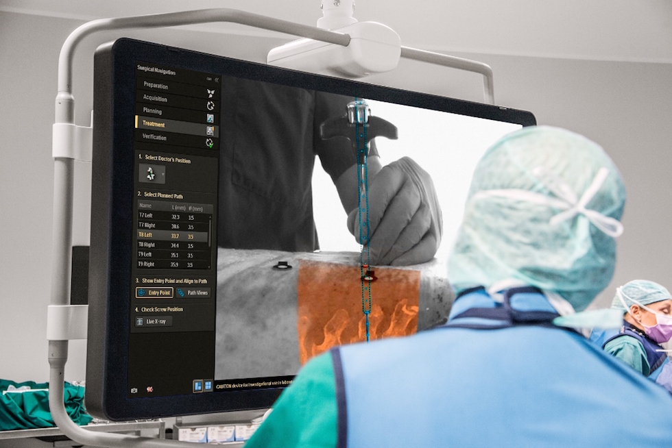 The system uses high-resolution optical cameras mounted on a flat-panel X-ray detector to image the surface of the patient. It then combines the external view captured by the cameras with the 3D internal view acquired by the X-ray system to create accurate real-time, augmented-reality images of the patient’s anatomy.
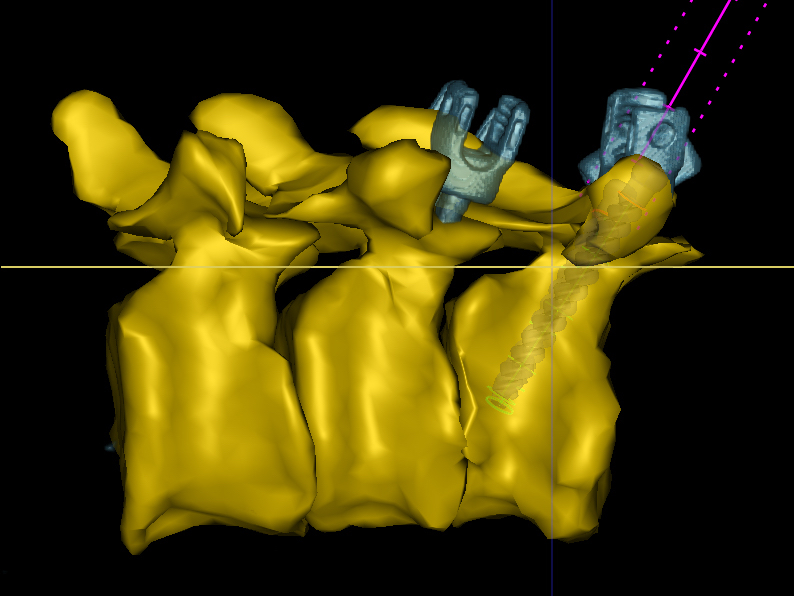
“We can make 3D acquisitions of the vertebrae of the spine, and show where they lie in the body, and we can also register what happens above the skin,” said Tabaksblat. “So now if the surgeon holds an instrument above the skin, we can show them a virtual path along which the instrument should be inserted into the body.”
The system generates different 3D views of the patient, from above and to the side, allowing the surgeon to align their instruments accurately before insertion.
The system has undergone pre-clinical trials in an experimental setting at Karolinska University Hospital in Stockholm, Sweden, and the Cincinnati Children’s Hospital Medical Center in the US. In the trials, which were recently published in the journal Spine, the technology was shown to improve the overall accuracy of screw placement from 64 per cent to 84 per cent.
See the full story here: https://www.theengineer.co.uk/augmented-reality-guides-surgeons-during-spine-cranial-and-trauma-surgery/