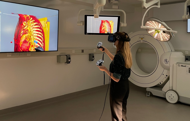
New Virtual Reality technology to repair hearts
The technology, which has been developed by researchers at Evelina London Children’s Hospital and King’s College London, brings together scans that are routinely used to plan congenital heart disease surgery to create a three-dimensional, beating digital double of the heart.
The researchers hope that using VR to plan and practice procedures will shorten operating times and reduce the need for multiple surgeries, leading to better outcomes and experiences for patients and their families. They hope that it could be in regular use within the next two years.
Trials of an early version of the technology, which used only echocardiograms (ultrasound scans of the heart) to create the VR heart, found that surgeons preferred it for understanding the anatomy of their patient’s hearts. They also reported that it increased their confidence and improved their decision making.
See the full story here: https://www.bhf.org.uk/what-we-do/news-from-the-bhf/news-archive/2022/february/new-virtual-reality-technology-to-repair-hearts